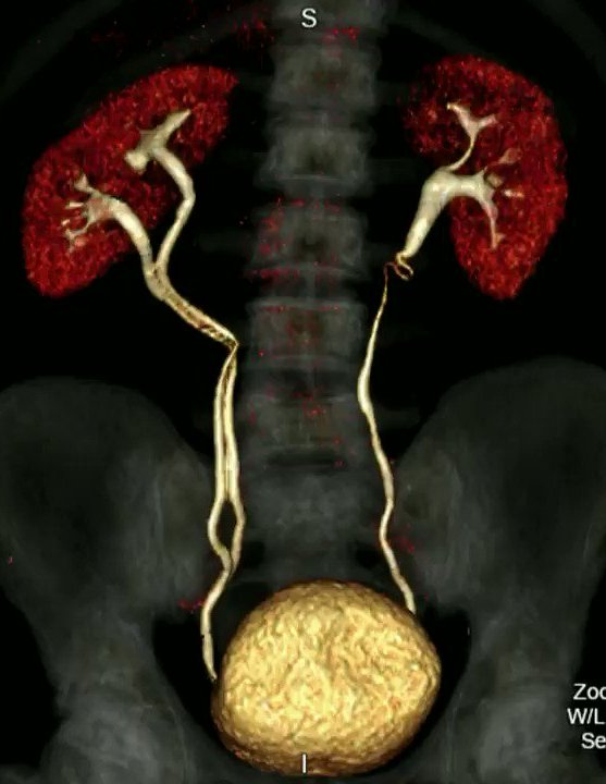
CT Urography
A computerized tomography (CT) urogram is an imaging exam used to evaluate your urinary tract, including your kidneys, your bladder and the tubes (ureters) that carry urine from your kidneys to your bladder.
CT urography uses X-rays to generate multiple images of a slice of the area in your body being studied, including bones, soft tissues and blood vessels. These images are then sent to a computer and quickly reconstructed into very detailed, 2-D and 3-D images.
During a CT urogram, an X-ray dye (iodine contrast solution) is injected into a vein in your hand or arm. The dye flows into your kidneys, ureters and bladder, outlining each of these structures. X-ray pictures are taken at specific times during the exam, so your doctor can clearly see your urinary tract and assess how well it’s working or look for any abnormalities.
A CT urogram may be used to help diagnose conditions that affect the urinary tract, such as:
- Kidney stones
- Bladder stones
- Infection
- Tumors or cysts
- Cancer
- Structural abnormalities
Preparation
Before a CT urogram, tell your doctor if you:
- Have any allergies, particularly to iodine
- Are pregnant or think you might be pregnant
- Have had a previous severe reaction to X-ray dyes
- Are taking any medications, such as metformin, nonsteroidal anti-inflammatory drugs (NSAIDS), anti-rejection drugs or antibiotics
- Have had a recent illness
- Have a medical condition, including heart disease, asthma, diabetes, kidney disease or a prior organ transplantation
In order to expand (distend) your bladder, you may be asked to drink water before a CT urogram and not to urinate until after the procedure. However, depending on your condition, guidelines about eating and drinking before your CT urogram may vary.